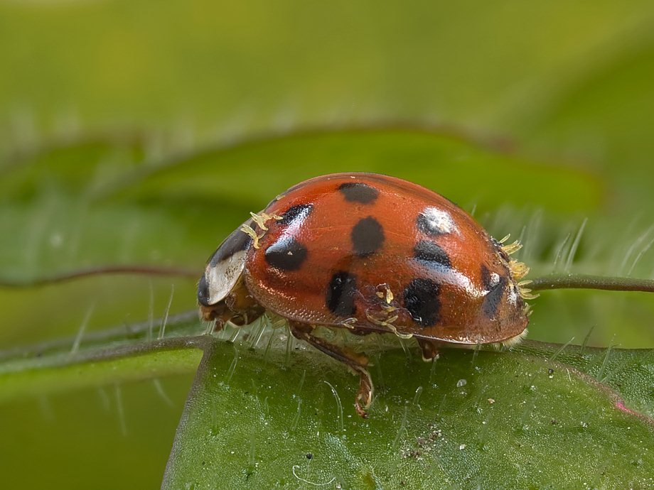
Microscopic fungal parasites reveal host’s behavior
Mycologists have long debated about which organisms should be or should be not counted as fungi. The Kingdom Fungi includes a lot of biodiversity; an estimate of 5.1 million species has been suggested, comprising all sorts of things—molds, the well-known mushrooms, plant and insect parasites, polypores, and some important model organisms such as Saccharomyces cerevisiae. To date, however, most fungi remain unknown and uncharacterized. The Laboulbeniales are perhaps (probably!) the most intriguing and yet the least studied of all insect-associated parasitic fungi.
Laboulbeniales?
The order Laboulbeniales (Fungi, Ascomycota) consists of obligate parasites living attached to the exterior of their invertebrate hosts, mainly beetles. Unlike the well-known mushroom structure (stem, cap + gills or pores), Laboulbeniales are microscopic organisms, referred to as thalli, of 0.15–1 mm in length, rarely more, bearing antheridia and perithecia on a receptacle with appendages.

These fungi exhibit great host specificity (i.e. one particular species lives on a single or a few host species), and are often remarkably adapted to a given position on the host body. This is important for the rest of this post: the occurrence of Laboulbeniales species on a precise portion of beetle integument is called “position specificity”.
Successful establishment of the parasite requires both the presence of a suitable host and favorable environmental conditions for the fungus. They produce sticky spores that are exclusively spread by the activities of the host, have a short life span, and cannot spread through air. Infection with Laboulbeniales through sexual contact is the most important type of direct transmission, it can therefore be referred to as a sexually transmitted disease. In ladybirds, Laboulbeniales are also socially transmitted as transmission also occurs when large numbers of lady beetles form aggregations in winter-time shelters. In spite of their parasitic nature, most Laboulbeniales are avirulent and seem to have little to no effect on the reproduction and survival of their host.
The order currently counts 145 genera and some 2,325 species, 1,260 of which have been described by one single person: Roland Thaxter (1858-1932), who did his research at the Farlow Herbarium (of the Harvard University Herbaria), as I did during my time as a PhD student.
Understanding beetle behavior
In research, even fundamental, we always want to answer some big questions. Now what can be possibly be answered using Laboulbeniales, tiny parasites without any harmful effects on their hosts? Tough question, but Lauren Goldmann seems to have found an interesting way of applying these fungi: using the specific positions of different species on their host in order to explain host beetle (mating) behavior. That is quite something. Let’s have a closer look.
Thirteen (!) species of the genus Chitonomyces have been described on the aquatic beetle Laccophilus maculosus (Coleoptera, Dytiscidae). Goldmann and Weir have shown by using molecular data that these 13 species reported to exhibit position specificity on L. maculosus were …
… placed neatly into pairs of morphotypes, resulting in synonymies and recognition of six phylogenetic species (one species is a triplet).
Goldmann M, Weir A. 2012. Position specificity in Chitonomyces (Ascomycota, Laboulbeniomycetes) on Laccophilus (Coleoptera, Dytiscidae): a molecular approach resolves a century-old debate. Mycologia 104(5): 1143-1158.
Moreover:
Male beetles are susceptible to all 13 morphotypes, opposed to only six morphotypes known on females.
Goldmann M, Weir A. 2012. Position specificity in Chitonomyces (Ascomycota, Laboulbeniomycetes) on Laccophilus (Coleoptera, Dytiscidae): a molecular approach resolves a century-old debate. Mycologia 104(5): 1143-1158.
For example, the (new) phylogenetic species Chitonomyces paradoxus (Peyr.) Thaxt. consists of the morphotypes Ch. unciger (Thaxt.) Thaxt. and Ch. paradoxes (Peyr.) Thaxt. The latter name has been kept because it is the oldest of both:
Chitonomyces paradoxus (Peyr.) Thaxt., Mem. Am. Acad. Arts Sci. 12: 287 (1896).
= Heimatomyces paradoxus Peyr., Sitzungsber. Kaiserl. Akad. Wiss., Math-Naturwiss. Cl., Abt. 1 [Wien] 64: 251 (1873).
= Chitonomyces unciger (Thaxt.) Thaxt., Mem. Am. Acad. Arts Sci. 12: 288 (1896).
= Heimatomyces unciger Thaxt., Proc. Am. Acad. Arts Sci. 30: 478 (1895).
Thalli of the Ch. paradoxus morphotype are positioned on the edge of the left elytron of both male and female Laccophilus maculosus. Chitonomyces unciger always parasitizes the left posterior claw of only males.

This rings a bell, doesn’t it? Well, it certainly did for the team from the State University of New York College of Environmental Science and Forestry. They found that the six phylogenetic species each comprise a pair – and in one case three – previously described morphological species that correspond to points of contact between male and female beetles during copulation. Bang, quite some results!

And this is not all. When placing the copulating male beetle in parallel orientation with the female, the positions of the morphotypes in males and females misalign (fig. A). It is shown that males are oriented in a diagonal (as in fig. B above), resulting in the alignment of all positions. This diagonal position is suggested to be demonstrated by males when females try to break free from the male hold, thereby increasing the overall pressure on the female. This specific pressure will result in the release of ascospores from the perithecium.
So, then why are thalli of Chitonomyces paradoxus present on the edge of the left elytron present on both males and females? Shoudn’t they just be present on females in that position? The funny thing about these beetles is that the male individuals just try to copulate with all, even with other males. Therefore, males are infected at both positions, because of the presence of so-called same-sex mounting behavior, which has never been observed in females. [Remember that the pairing morphotype, Ch. unciger, is positioned at the left posterior claw of only males.]
End of story, problem solved, and one very nice application for my beloved organisms.
References
Haelewaters D, Dima B, Abdel-Hafiz BII, Abdel-Wahab MA, Abul-Ezz SR, Acar I, Aguirre-Acosta E, Aime MC, Aldemir S, Ali M, Ayala-Vásquez O, Bakhit MS, Bashir H, Battistin E, Bendiksen E, Castro-Rivera R, Çolak ÖF, De Kesel A, de la Fuente JI, Dizkırıcı A, Hussain S, Jansen GM, Kaygusuz O, Khalid AN, Khan J, Kiyashko AA, Larsson E, Martínez-González CR, Morozova OV, Niazi AR, Noordeloos ME, Pham THG, Popov ES, Psurtseva NV, Schoutteten N, Sher H, Türkekul İ, Verbeken A, Ahmad H, Afshan NS, Christe P, Fiaz M, Glaizot O, Liu J, Majeed J, Markotter W, Nagy A, Nawaz H, Papp V, Péter Á, Pfliegler WP, Qasim T, Riaz M, Sándor AD, Szentiványi T, Voglmayr H, Yousaf N, Krisai-Greilhuber I. 2020. Fungal Systematics and Evolution 6. Sydowia 72: 231-356. https://doi.org/10.12905/0380.sydowia72-2020-0231 [pdf]
Goldmann LM, Weir A. 2012. Position specificity in Chitonomyces (Ascomycota, Laboulbeniomycetes) on Laccophilus (Coleoptera, Dytiscidae): a molecular approach resolves a century-old debate. Mycologia 104(5): 1143-1158. https://doi.org/10.3852/11-358 [pdf]
Rossi W, Santamaría S. 2012. Rodaucea, a new genus of the Laboulbeniales. Mycologia 104(3): 785-788. https://doi.org/10.3852/11-337
Blackwell M. 2011. The Fungi: 1, 2, 3… 5.1 million species? American Journal of Botany 98(3): 426-438. https://doi.org/10.3732/ajb.1000298
De Kesel A. 1996. Host specificity and habitat preference of Laboulbenia slackensis. Mycologia 88(4): 565-573. https://doi.org/10.1080/00275514.1996.12026687
Weir A, Beakes GW. 1995. An introduction to the Laboulbeniales: a fascinating group of entomogenous fungi. Mycologist 9(1): 6-10. https://doi.org/10.1016/S0269-915X(09)80238-3
Tavares II. 1985. Mycologia Memoir 9: 1-627.
Thaxter R. 1896. Contribution towards a monograph of the Laboulbeniaceae. Memoirs of the American Academy of Arts and Sciences 12(3): 187-429.


Very interesting read. Any idea why the parasites were located at copulation sites though? Do they feed (albeit not exclusively) on the beetles’ ejaculate?
The parasites are located at various positions on the integument, where male and female make contact.
For example, there’s a morphotype at the left posterior claw of only males (Chitonomyces unciger). The morphotype that forms a pair with this one, i.e. Chitonomyces paradoxus (= also the name for the phylogenetic species), is positioned only at the edge of the left elytron of both males and females. Males try to hold females during copulation with primarily the posterior legs, so this makes sense for the positioning of the both morphotypes at males and females. Males are infected at both positions [left posterior claw + edge of left elytron] because of the presence of self-infection and “same-sex mounting behavior”, which has never been observed in females.
I see, but what do the parasites get out of the positioning?
Hmm, I don’t think I completely understand your question here. :-/
The only way of transmission of Laboulbeniales is by ascospores, which seem to be released primarily by pressure of the male hold. Since the spores cannot spread through air or water, they need to be delivered precisely. That is, I guess, why this “position specificity” phenomenon is so important in Laboulbeniales. Same-sex and opposite-sex mating behavior will position the ascospores in a precise portion of the new host’s integument – no way around it.
Taking Laccophilus maculosus as example, only 13 precisely delineated spots will be infected by Laboulbeniales (6 phylogenetic species, 13 morphotypes), full stop.
Great blog post.